Sparta, a 9-year-old neutered male domestic shorthair cat, is presented in lateral recumbency for weakness and a wobbly gait of 2-days’ duration. His owner states Sparta started showing a nonspecific decrease in mental alertness, appearing lethargic, and exhibiting uncharacteristic behaviors the week prior to onset of weakness but had been previously healthy.
History
Sparta is an indoor-only cat. Another indoor-only cat also lives in the household. Both cats were acquired as kittens and have been housed together for >5 years. Vaccinations and routine monthly preventives are current in both cats, and both cats are free fed a commercial dry cat food.
Appetite, fluid intake, urination, and defecation have been normal, but energy and activity have been reduced. There is no history of neurologic or orthopedic disorders.
Physical Examination
On physical examination, Sparta is unable to stand unassisted and shows persistent cervical ventroflexion with the head held low. His temperature is 98.6°F (37°C), heart rate is 144 bpm, and respiratory rate is 30 breaths per minute. Mentation is determined to be depressed.
Hydration is judged to be adequate, and the urinary bladder is distended. The remainder of the physical and neurologic examinations are unremarkable except for mild discomfort elicited with palpation of larger muscles (eg, pelvic and proximal rear limb musculature) and several small retinal hemorrhages. Systolic blood pressure via Doppler in the thoracic limb is 200 mm Hg.
Investigation
Results of laboratory analysis (Table) show hypokalemia and markedly elevated creatine kinase. Abdominal radiographs reveal nephroliths in both kidneys, several small cystoliths, and a soft tissue mass cranial to the right kidney.
Table 1: Clinicopathology Results Obtained at Presentation*
*Abnormal values are in bold.
How would you diagnose and treat this patient?
Treatment & Outcome
Radiographic findings were an indication for advanced imaging of the abdomen. Based on the available modalities, ultrasonography was elected so the procedure could be performed promptly without placing the patient under general anesthesia. Ultrasound results showed an enlarged right adrenal gland (Figure).
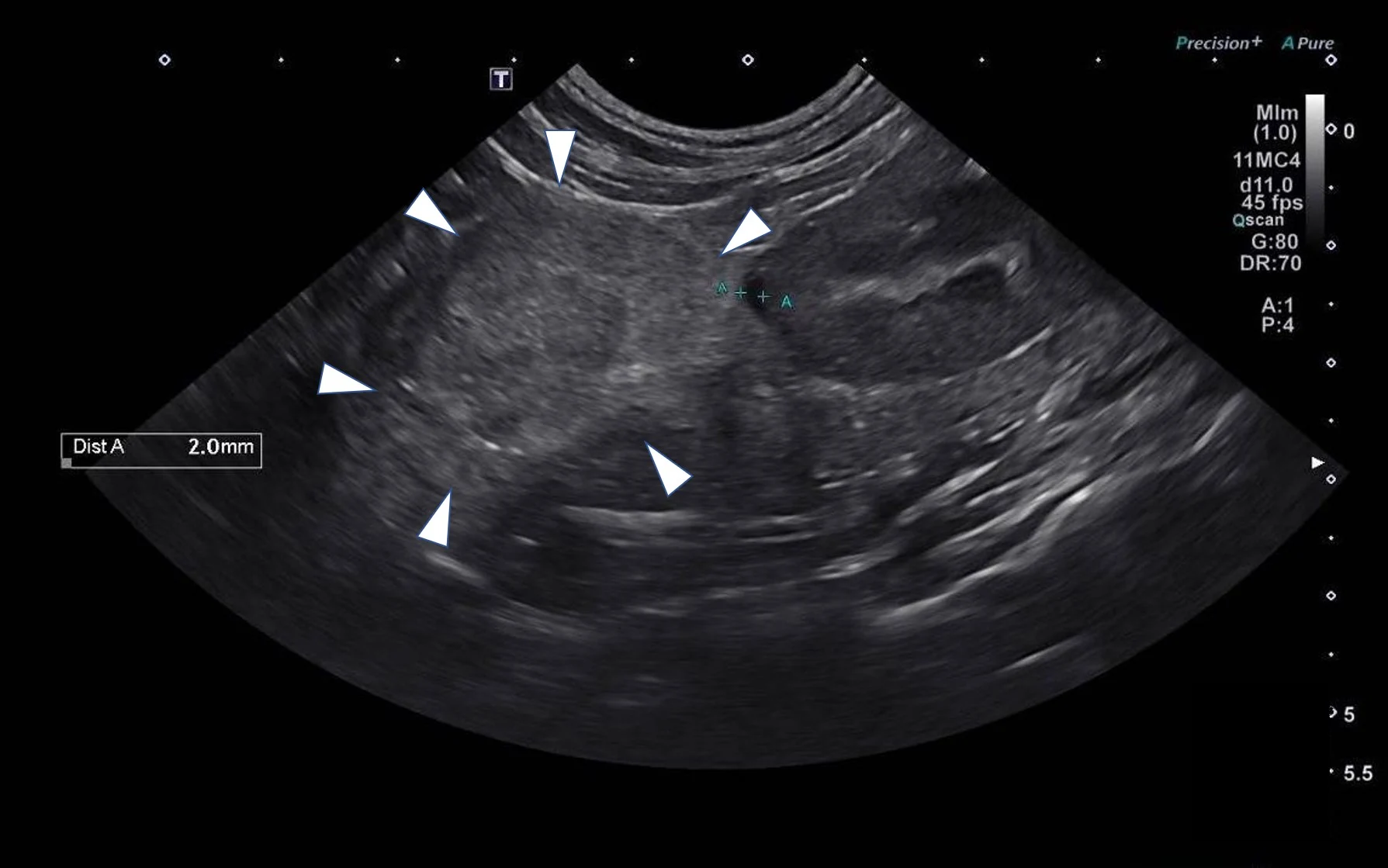
Abdominal ultrasound showing a 4.2- × 3-cm mass involving the right adrenal gland (arrowheads)
Ultrasound findings, along with other clinical and laboratory results, suggested Sparta had an aldosterone-producing adrenal tumor causing primary hyperaldosteronism.1 Hyperaldosteronism is characterized by hypokalemia (secondary to potassium-wasting effects of aldosterone) and severe hypertension (secondary to sodium-mediated volume expansion).1,2 Serum aldosterone was subsequently measured from a sample collected at presentation and found to be markedly elevated (>187.4 ng/dL; reference interval, 7-14 ng/dL).
While waiting for the serum aldosterone result to confirm the diagnosis, Sparta was hospitalized to address aldosterone-mediated total body potassium depletion, which clinically manifests as serum hypokalemia. Potassium, as potassium chloride added to lactated Ringer’s solution, was administered IV until the serum potassium level increased and stabilized. Serum potassium was maintained between 2.6 to 2.8 mEq/L while IV supplementation was administered. This clinical improvement allowed transition to an oral medication.
Sparta was discharged and therapy continued in the home with potassium gluconate (3 mEq PO every 12 hours) added to the diet to increase potassium intake.3 Spironolactone (12.5 mg [≈2 mg/kg] PO every 12 hours), a potassium-sparing diuretic, was also initiated to address suspected aldosterone-mediated hypokalemia and hypervolemia.3 Adrenalectomy was indicated,1-3 but the owner declined surgery.
The case was lost to follow-up soon after discharge.