When T4 Values are Normal

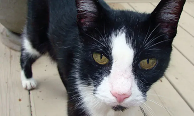
Socks, a 15-year-old, castrated, domestic shorthaired cat, presented for anorexia, vomiting, and weight loss.
HISTORYThe owner reported weight loss of 9 months’ duration and a 3-day history of anorexia, lethargy, and vomiting (yellow fluid) once or twice daily. Socks’ appetite and activity level had previously been normal.
T4 concentrations can fluctuate throughout the day, even in normal cats.
According to the referring veterinarian, a serum biochemistry profile revealed elevated alanine transaminase (ALT) and blood urea nitrogen (BUN), although the actual values were not specified. Socks was treated with amoxicillin/clavulanic acid at 62.5 mg PO Q 12 H before referral.
PHYSICAL EXAMINATIONOn presentation for referral examination, Socks appeared agitated, with a rectal temperature of 99.2°F; pulse rate, 144 beats/min; and respiratory rate, 30 breaths/min. Socks weighed 3.6 kg and had a body condition score of 3/9 with a generalized decrease in muscle mass. He had a poor hair coat, dry mucous membranes, slightly increased skin turgor, and estimated dehydration of 5%.
There was no abdominal pain or evidence of masses or organomegaly on palpation. A thyroid nodule (ie, “thyroid slip”) was noted on palpation of Socks’ neck. No murmur or arrhythmia was noted on auscultation of his heart.
ALT = alanine transaminase, BUN = blood urea nitrogen
LABORATORY RESULTSA complete blood count (CBC) showed a stress leukogram and increased packed cell volume (PCV) of 53%. A serum biochemistry profile revealed increased concentrations of BUN (48 mg/dL; range, 10–40 mg/dL), ALT (127 U/L; range, 7–60 U/L), and globulin (6.6 g/dL; range, 4.1–6 g/dL). Serum creatinine was within reference range (1.9 mg/dL; range, 0.4–2 mg/dL). Urinalysis revealed pyuria and bacteriuria, and the specific gravity was 1.034. A postantibiotic urine culture (urine obtained by cystocentesis) was negative.
Total thyroxine (T4) was 3.1 mcg/dL (range, 0.8–4 mcg/dL) and serum creatine kinase was 111 U/L (range, 50–225 U/L), ruling out myopathy as the cause of increased ALT.
Patients with mild hyperthyroidism or concurrent illness can have total T4 values in the upper half of the normal range.
Because of Socks’ acute exacerbation of clinical signs, the owners elected to pursue additional diagnostics to help rule out other differentials. Thoracic radiographs revealed no abnormalities. Abdominal ultrasound revealed mixed echogenic debris in the urinary bladder. There was no evidence of pyelonephritis, and the liver appeared to be normal in size and echogenicity. Ultrasound-guided liver aspiration was nondiagnostic.
Diagnostic Differentials• Hyperthyroidism (primary differential)• Chronic renal failure• Primary hepatopathy or neoplasia• Lymphoma• Inflammatory bowel disease• Myopathy
INITIAL TREATMENTLower urinary tract infection was presumptively diagnosed based on pyuria and bacteriuria, despite the negative urine culture. Idiopathic cystitis was also possible but considered less likely due to the lack of lower urinary tract signs (eg, pollakiuria).
Supportive therapy was initiated, and Socks was given intravenous lactated Ringer’s solution overnight but switched to subcutaneous fluids because he removed the intravenous catheter. He also received amoxicillin/clavulanic acid at 50 mg PO Q 12 H. No further vomiting occurred, and Socks’ appetite returned. His temperament also improved and hydration status normalized.
ASK YOURSELF...Although Socks’ acute signs resolved with supportive therapy, the cause of chronic weight loss had not been identified. Which diagnostic procedure(s) would you conduct next?
A. Additional thyroid testing (eg, total T4, free T4 [fT4], triiodothyronine [T3] suppression tests)B. Endoscopy and gastric and duodenal biopsiesC. Abdominal exploratory and gastric and small intestinal biopsiesD. Workup for hepatobiliary disease
ALT = alanine transaminase, BUN = blood urea nitrogen, CBC = complete blood count, fT4 = free thyroxine, PCV = packed cell volume, T3 = triiodothyronine, T4 = thyroxine
CORRECT ANSWER:A. Additional thyroid testing
While the total T4 concentration is increased in most cats with hyperthyroidism, some patients with mild hyperthyroidism or concurrent illness have a total T4 value within the upper half of the reference range (>2 mcg/dL). One reason for this divergence is that the total T4 concentration can fluctuate throughout the day even in normal cats. Therefore, in a hyperthyroid cat, the T4 concentration may be within the reference range for part of the day, but when rechecked on a different day, above the reference range.
Sick euthyroid syndrome may also result in a total T4 concentration within reference range in hyperthyroid cats. Because fT4 (by equilibrium dialysis) concentrations are usually less affected by nonthyroidal illness, some hyperthyroid cats with a normal T4 concentration may have an increased fT4. Unfortunately, some studies have shown that fT4 concentrations may also be increased in euthyroid cats with nonthyroid illness (particularly chronic renal failure). Thus the specificity of a fT4 test may be less than that of the total T4 test for these patients, making the utility of fT4 results less straightforward. Assessing the hypothalamic–pituitary–thyroid axis with the T3 suppression test can help differentiate hyperthyroid cats from euthyroid cats in these situations.
Assessing the hypothalamic–pituitary–thyroid axis with a T3 suppression test can help differentiate hyperthyroid cats from euthyroid cats.
WEIGHING ALL CLINICAL SIGNSAlthough most untreated hyperthyroid cats are hyperactive and polyphagic, lethargy and anorexia are present in some patients (approximately 10%) and may be suggestive of concurrent disease. Other common causes of chronic weight loss in geriatric cats include chronic renal failure, lymphoma, and inflammatory bowel disease.
Physical examination findings, hemoconcentration, and increased BUN concentration were consistent with dehydration, which could be secondary to decreased intake and loss through vomiting. However, enhanced protein turnover can increase the BUN concentration in patients with hyperthyroidism independent of the hydration status. The high-normal creatinine concentration should also be viewed in light of the body condition score; the value may actually be above “normal” for a cat with muscle wasting.
Accurate assessment of renal dysfunction in hyperthyroid cats is not always straightforward. Since a dehydrated cat with normal renal function should have a urine specific gravity greater than 1.035, mild renal disease could not be ruled out completely in Socks. However, in light of his hemoconcentration and normal creatinine level, primary renal disease was considered to be a less likely cause of the increased BUN value.
In addition, pyelonephritis could not be ruled out based on ultrasonographic findings but would not be expected to cause chronic weight loss without abnormalities on ultrasound.
While increased ALT concentration is supportive of a diagnosis of hyperthyroidism, other differentials include primary hepatopathy and myopathy.
CONFIRMING HYPERTHYROIDISMFor Socks, recheck of the total T4 found a concentration still within normal range (2.2 mcg/dL). A T3 suppression test was conducted. Baseline T4 was 2.2 mcg/dL, and the post-T4 finding was 2.3 mcg/dL. Lack of T4 suppression by T3 confirmed the diagnosis of hyperthyroidism.
In this situation, a T3 suppression test was chosen due to concerns about the specificity of fT4 testing. However, fT4 measurement would also have been appropriate if interpreted accordingly. Ideally, these tests are conducted when the animal is clinically well, but this was not possible in this case.
Find More!
Read How to Refer: The Hyperthyroid Cat (April 2006) to learn more about when you might consider referring your hyperthyroid patients, plus Consultant on Call: Feline Hyperthyroidism (March 2006) for additional information on diagnosing and managing this disease process. Available at cliniciansbrief.com/journal.
Inducing a euthyroid state with methimazole and monitoring renal values are recommended for at least 1 month before starting I-131 therapy.
TREATMENTTreatment of hyperthyroidism decreases the glomerular filtration rate (GFR) and may unmask preexisting renal disease in cats, even if azotemia is not present when treatment is initiated. It is important to rehydrate dehydrated patients prior to initiating methimazole treatment. A conservative (low) dose should be used initially because these patients may be predisposed to dehydration; dehydration combined with decreased GFR could result in decreased renal perfusion and damage.
Although I-131 therapy could be considered in the future, inducing a euthyroid state with methimazole therapy and monitoring renal values are recommended for at least 1 month before initiating I-131 treatment to ensure that azotemia does not develop. Socks was started on 2.5 mg of methimazole PO Q 24 H. Treatment with amoxicillin/clavulanic acid (50 mg PO Q 12 H) was also continued for 2 weeks.
After 2 years of treatment with methimazole, Socks continued to do well, as demonstrated by his body condition, demeanor, and energy level.
OUTCOMESocks returned for recheck CBC, serum biochemistry profile, urinalysis, and urine culture 2 weeks after discharge. His owners reported a return to normal activity and appetite and no vomiting.
Laboratory findings were all within reference range, and the T4 was 3.0 mcg/dL. The owners were instructed to have the CBC, renal panel, and T4 rechecked by the referring veterinarian every 2 weeks for the first month, then every 6 months.
Socks has continued to do well for over 2 years. Based on T4 concentrations, his methimazole dose has been increased to 7.5 mg/day, but he has gained 1.7 kg and his renal parameters remain within reference range.
BUN = blood urea nitrogen, CBC = complete blood count, fT4 = free thyroxine, GFR = glomerular filtration rate, T3 = triiodothyronine, T4 = thyroxine
WHEN T4 VALUES ARE NORMAL • Patty Lathan
Suggested Reading
Interpretation of endocrine diagnostic test results for adrenal and thyroid disease. Kemppainen RJ, Behrend EN. In Bonagura J, Twedt D (eds): Kirk’sCurrent Veterinary Therapy XIV—Philadelphia: WB Saunders, 2008, pp 170-174.Survival and the development of azotemia after treatment of hyperthyroid cats. Williams TJ, Peak KJ, Brodbelt D, et al. J Vet Intern Med 24:863-869, 2010.Testing for hyperthyroidism in cats. Shiel RE, Mooney CT. Vet Clin North Am Small Anim Pract 37:671-691, 2007.