Ultrasonography at the Point of Care: Blue's Case
Sponsored by Butterfly
Ultrasonography in veterinary medicine has historically been reserved for use in referral practices, often performed by trained radiologists and other specialists for its diagnostic capabilities. As technology has advanced, ultrasound machines have become more accessible to general veterinary practitioners. These new point-of-care devices are generally portable, affordable, and reliable, providing an alternative to the traditional large machines.
Meet Blue
Blue, a 12-year-old neutered male domestic shorthair cat, was presented for gradual weight loss and declining appetite over the past several weeks. His owner reported occasional vomiting, but this was not new. Thirst, urination, and bowel movements were normal.
Blue had been examined annually for the last several years and had no previous history of abnormalities on blood work or physical examination. Blue was very stressed by travel, usually presenting to the clinic panting. Gabapentin typically helped him, but his owner preferred to minimize the number of trips to the veterinary clinic to decrease stress and liked to accomplish as much as possible in a single visit.
Physical examination showed a 4-lb weight loss and moderate muscle loss. A palpable mid-abdominal mass was also noted. The examination was otherwise unremarkable.
The owner approved blood work and radiography. Mild liver enzyme elevations and moderate lymphocytosis were identified on blood work. Radiography showed mild hepatomegaly, dorsal displacement of the stomach, and a mass effect in the mid-abdomen. Based on these results, abdominal ultrasonography was recommended. Blue's owner agreed but was concerned about him tolerating the ultrasound while away from her.
What Is the Butterfly iQ+ Vet?
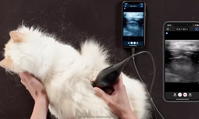
Butterfly iQ+ Vet is a point-of-care ultrasound probe featuring curvilinear, linear, and sector array formats in a single device that readily connects to a compatible cell phone and/or tablet through an application. It requires minimal storage space and is highly portable, allowing patient-side ultrasonography to be performed in the examination room with the owner.
The Butterfly iQ+ Vet utilizes Ultrasound-on-Chip™ technology and features veterinary-specific presets to simplify and optimize format choices, which include depth, frequency, and gain, for maximum image quality.
The Butterfly iQ+ Vet utilizes Ultrasound-on-Chip™ technology and features veterinary-specific presets to simplify and optimize format choices, which include depth, frequency, and gain, for maximum image quality. Doppler, M-mode, and other actions and tools are available with an intuitive touchscreen interface. The program integrates with a clinic’s existing imaging system and provides cloud-based image storage.
Butterfly iQ+ Vet also has TeleGuidance™*, which allows the tableside operator to collaborate with a trained peer who can remotely view images in real time, see how the probe is positioned on the patient through the cell phone’s camera feed, and adjust settings. This service can work between any colleagues with compatible devices and software so a relationship with a specialist of the veterinarian’s choosing can be established.
Back to the Case
In Blue’s case, a circumferential intestinal mass was identified in the small intestinal tract. The mesenteric lymph nodes were enlarged, and the liver had abnormal echogenicity. Referral to a specialist was offered, and aspiration of the intestinal mass and liver were discussed and performed at the primary care facility.
With the patient having been fasted that morning, ultrasound-guided aspiration was performed during the appointment with the Butterfly iQ+ Vet. Needle Viz™ technology allowed for easy visualization of the needle to facilitate safe aspiration of the intestinal mass. Aspirates of the liver were collected as well, and all samples were sent to a clinical pathologist for analysis.
Aspiration results were consistent with lymphoma in both the intestine and liver. Referral to an oncologist was provided to initiate treatment.
Conclusion
Having in-clinic, patient-side ultrasonography allows for accessible diagnostic imaging and improved peace of mind for the pet owner. With image visualization done through an application on a handheld device, collaboration with peers for support during image collection and image interpretation is also possible, empowering the general practitioner and expanding the number of patients who can benefit from the capabilities of ultrasonography.
For trained veterinary professionals only.
*TeleGuidance™ is only available for compatible iOS devices.