Diagnosis of Demodicosis in Dogs & Cats
This article is the first of 2 articles on demodicosis in dogs and cats—this first article will cover diagnosis of demodicosis; the second will address treatment, including medications, follow-up, complications, and prognosis.
PROFILE
Definition
Demodicosis occurs when Demodex mites, which are part of an animal’s normal flora, proliferate in the skin (most often the hair follicle).
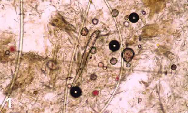
Figure 1 (above): Long-bodied canine Demodex mite (original magnification, 1000x)
Causes
Three species of Demodex mites affect dogs: D canis, D injai (long-bodied mite; Figure 1), and D cornei (short-bodied mite).<sup1, 2sup>
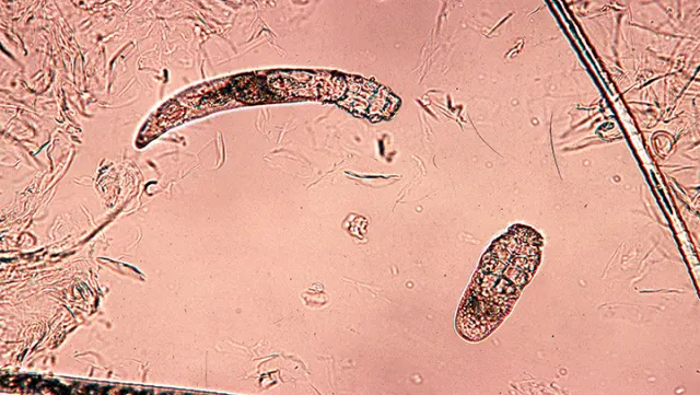
Two species affect cats: D cati (long-bodied mite) and D gatoi (short- and wide-bodied mite; Figure 2).3
Feline Demodex mites: D cati and D gatoi (original magnification, 40x)
Signalment
Demodicosis can develop at any age.
No sex predilection is seen in dogs or cats.
Breeds at highest risk for developing juvenile-onset generalized demodicosis due to D canis include shar-peis, pit bulls, Boston terriers, English bulldogs, boxers, miniature pinschers, Great Danes, and pugs.4
Emerging evidence suggests that wire-haired fox terriers, and terriers in general, may also be predisposed to demodicosis.4-6
There is no recognized breed predilection in cats.
Risk Factors
Risk factors that affect both dogs and cats include chronic severe disease states, neoplasia, long-term glucocorticoid use, and chemotherapy.
In dogs, risk factors for juvenile-onset generalized demodicosis include pyoderma, coccidiosis, hookworms, short hair coat, and lack of participation in a preventive care wellness plan.4
Focal demodicosis is common in puppies, and physiologic stress and debilitation are risk factors.
In cats, D gatoi is contagious. Cats at risk for this infestation tend to come from high-density populations.
Related Article: Treatment of Demodicosis in Dogs & Cats
Pathophysiology
Clinical disease results from overproliferation of mites in the skin due to defects or compromise of the skin’s immune system.
Canine demodicosis can mimic any skin disease; the rule of thumb is "demodicosis until proven otherwise."
Studies using dog leukocyte antigen class II have identified common markers in young dogs with generalized demodicosis, suggesting that this antigen may be an important immunologic risk factor for the disease in dogs.7
Demodex mites found on skin scrapings, plucked hair, ear swabs, and fecal samples are considered clinically significant when interpreted in conjunction with typical clinical signs.8
Diagnosis
View a Table for a description of clinical signs of demodicosis in dogs and cats
Definitive Diagnosis
Dogs
Any finding of mites indicates demodicosis.5,6 Mixed infestations of D canis and D injai can occur.
Mites are found on deep-skin scrapings or via hair trichograms; the latter are useful for sampling sites that are close to the eyes or difficult to scrape (eg, interdigital areas).
Hair plucking may be the diagnostic test of choice for sampling dogs with greasy hair.5,6
Cats
With otic demodicosis, mites are found on mineral oil cytologic testing of ear exudates.
D cati mites tend to be easily found on skin scrapings.
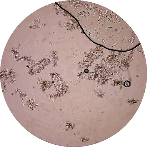
D gatoi mites can be difficult to find even when the patient is severely pruritic. For these mites, suggested tests include wide superficial skin scrapings, hair plucking, fecal flotations (Figure 3), or response to therapy.
Demodex gatoi mites found on fecal sample from cat with pruritis, self-trauma, and hair loss on ventral abdomen (original magnification, 400x)
Differential Diagnosis
Dogs: Canine demodicosis can mimic any skin disease; the rule of thumb is “demodicosis until proven otherwise.”
Cats: Consider demodicosis in any cat with hair loss, symmetric alopecia, or pruritus.
Laboratory Testing
Bacterial cultures of skin: Should be performed if deep pyoderma is present or if there is a history of glucocorticoid use or long-term antibiotic therapy (dogs)
Complete blood count or serum biochemical profile: In dogs with deep pyoderma that may be septic or dehydrated
Laboratory testing is most helpful when searching for the underlying cause of adult-onset demodicosis in dogs.
Fecal flotation examination: In pruritic cats to identify D gatoi
Genetic testing for ABCB1-δ genotype: To screen for drug sensitivity to avermectins in breed-sensitive dogs (eg, herding dogs, sight hounds); recommended for dogs with severe generalized demodicosis3
Impression smears: To diagnose concurrent microbial overgrowth in dogs and cats
Additional Laboratory Testing
Except for fecal flotation examinations in cats, laboratory testing is most helpful when searching for the underlying cause of adult-onset demodicosis in dogs or a medical condition associated with D cati in an adult cat. Testing may include but is not limited to:
Blood smear evaluation
Complete blood count
Fecal flotation
Infectious disease titers
Radiography of thorax and abdomen
Retroviral screening
Serum biochemical analysis
Urinalysis
Fine-needle aspirates of lymph nodes may contain mites.
Skin biopsy may be helpful in shar-pei dogs and dogs with severe pododermatitis.