Anesthesia in Hepatic Disease
Khursheed Mama, DVM, DACVAA, Colorado State University
Marlis Rezende, DVM, PhD, DACVAA, Colorado State University
You have asked…
What are general considerations regarding anesthesia for a patient with hepatic disease?
The expert says…
The liver has a number of important roles in the body: It secretes bile, filters toxins from blood, and is responsible for the metabolism, biotransformation, and elimination of endogenous and exogenous compounds (including many anesthetic drugs).
The choice of anesthetic drugs for a patient with hepatic disease depends on the type and severity of disease.
Key functions of the liver include synthesis and metabolism of carbohydrates (glucose production and glycogen storage), lipids (production of energy, triglycerides, and cholesterol), and proteins (production of albumin and coagulation factors and conversion of ammonia into urea). These functions are facilitated by the dual blood supply (providing nutrients and removing toxins), with about 70% of liver blood flow originating from the digestive tract via the portal vein and the remainder from the hepatic artery.1
Laboratory Values in Liver Disease
Because the liver’s many functions, signs and laboratory values vary with the severity, duration (acute or chronic), and location (parenchymal or cholestatic) of disease. Patients with liver disease can present from clinically normal to significantly debilitated. In extreme cases, neurologic signs, including seizures, may be observed.
Elevations in liver enzymes suggest damage to hepatocytes (alanine aminotransferase [ALT] and aspartate aminotransferase [AST]) or biliary obstruction (alkaline phosphatase [ALP]), if other causes such as muscle trauma or bone disease can be ruled out. Increased gamma–glutamyl transpeptidase (GGT) values further support the diagnosis of hepatic disease. Albumin, glucose, blood urea nitrogen, and cholesterol values may be low in patients with liver failure.
Reduced production of coagulation factors can lead to bleeding disorders. Similarly, ascites may be evident in a patient with severe hypoalbuminemia or portal hypertension. Evaluation of direct and indirect bilirubin, pre- and postprandial bile acids, and ammonia levels can also be used to further characterize the disease process.
In addition to a thorough physical examination, evaluation of the laboratory parameters discussed above is essential before developing an anesthesia plan. The focus is not only on anesthesia drug selection but also on treating or compensating for sequelae of the primary disease.
Related Article: Anesthesia for Pancreatic Disease
Administering Fluids
Patients presented with hypoglycemia should receive dextrose supplementation, and blood glucose values should be monitored. At fluid rates commonly used during anesthesia (5–10 mL/kg/h), dextrose may be added to isotonic (replacement) IV fluids to achieve concentrations of 1%–2.5%. Colloids may be necessary in hypoalbuminemic patients, with the type (synthetic vs fresh or fresh–frozen plasma) determined based on coagulation status. The authors commonly use 2 mL/kg/h of hetastarch during the anesthetic period in patients with low albumin and normal coagulation parameters (with a limit of about 20 mL/kg q24h; lower in patients with impaired coagulation).
Isotonic fluid rates are typically decreased to 3–5 mL/kg/h in hypoalbuminemic patients. While lactate-containing solutions (eg, lactated Ringer’s solution) can be used safely in most patients during anesthesia, acetate- or gluconate-containing solutions (eg, Plasma-Lyte [abbottanimalhealth.com], Normosol [hospira.com]) may be preferred in severely compromised patients, as lactate requires liver metabolism. In patients presented with ascites, abdominal fluid may need to be drained before anesthesia administration to reduce intraabdominal pressure and facilitate circulation and ventilation.
Patients with high ammonia and serum bile acid levels, as well as neurologic signs, may be especially challenging to manage. Appropriate therapy to reduce ammonia levels should be instituted before anesthetic induction to reduce the risk for hepatic encephalopathy and seizures postanesthesia.
Avoiding Hemorrhage
In addition to the potential for bleeding resulting from a reduction in coagulation factors, there is significant potential for hemorrhage when working in the area of the liver. Crystalloids and synthetic colloids may be appropriate for initial treatment, but it is prudent to have patients typed and cross-matched in the event that other blood products (eg, packed RBCs, whole blood) are needed. Fresh or fresh–frozen plasma may be administered without typing to provide coagulation factors unless the animal has a history of transfusion reactions. Placement of a second IV catheter in high-risk animals is recommended to facilitate rapid IV fluid administration to maintain oxygen delivery.
IV volume expansion will help maintain circulating blood volume and adequate blood pressure, which are essential to supporting organ function and particularly important when dealing with compromised tissue. Direct blood pressure monitoring is recommended in compromised animals or those at increased risk for surgical complications. Inotropic drugs (eg, dopamine or dobutamine, 2–7 µg/kg/min) should also be available in case of hypotension that is refractory to treatment with intravenous volume replacement and decreasing anesthetic dose.
Vasopressors (eg, norepinephrine, phenylephrine, vasopressin) are used occasionally in severely compromised patients that are unresponsive to intravenous fluid resuscitation and inotropes.
Metabolic acidosis and electrolyte imbalances (eg, hypocalcemia, hypokalemia) may be present in severe cases. These changes can negatively affect cardiovascular function (and impair the response to inotropic drugs) and so should be corrected. The authors advise that anesthesia textbooks should be consulted for dose calculations and administration rates.
Related Article: Geriatric Anesthesia & Analgesia
Maintaining Oxygenation & Ventilation
Adequate oxygenation and ventilation should be maintained throughout the perioperative period. Oxygen administration before anesthetic induction and during the recovery period is recommended to prevent hypoxemia. Mechanical ventilation should be used to keep arterial carbon dioxide levels within the normal range, as both hyper- and hypocapnia have been shown to have negative effects on hepatic blood flow.
Monitoring
The degree of monitoring required depends on the severity of hepatic disease but should include, at a minimum, blood pressure, heart rate and rhythm, oxygenation, ventilation, body temperature, and blood glucose concentrations. In compromised animals, direct arterial blood pressure monitoring is recommended and additional monitoring of blood gases, electrolytes, acid–base status, packed cell volume, total protein, and coagulation status are important to facilitate timely intervention. Supportive therapy should be tailored to each animal’s needs and is essential to improve outcome in animals with hepatic disease.
Developing an Anesthesia Plan
The choice of anesthetic drugs for a patient with hepatic disease depends on the type and severity of disease. In general, preference should be given to short-acting and/or reversible drugs that do not depend exclusively on the liver for metabolism. In addition, these drugs should preserve cardiovascular stability or at least cause minimal depression.
Opioids (eg, hydromorphone or oxymorphone, 0.05–0.1 mg/kg SC or IM) may be used in dogs to provide sedation and analgesia and facilitate a dose reduction of anesthetic induction and maintenance agents. Opioids are reversible and cause minimal cardiovascular depression, except for bradycardia, which can be prevented or treated with an anticholinergic agent (eg, atropine, 0.02–0.04 mg/kg SC or IM; glycopyrrolate, 0.01–0.02 mg/kg IM). Profound sedation can be observed in patients with severely compromised liver function; lower doses might be preferable in these animals. Opioids may cause spasm of the sphincter of Oddi, but this effect can be countered with the use of anticholinergic drugs.Sedatives & Anesthetics
Acepromazine and α2-agonists are generally avoided because of their negative cardiovascular effects. Acepromazine, which has a long duration of action, depends exclusively on the liver for clearance (not reversible) and causes vasodilation and secondary hypotension. While α2-agonists (eg, dexmedetomidine) are reversible, they cause a marked decrease in cardiac output and hepatic blood flow.
Induction Agents
Induction drugs should always be titrated to effect in order to minimize cardiovascular depression, especially in patients with hypoproteinemia and acidemia, as these patients are more likely to experience more profound effects because of increased availability of the unbound (active) drug.
In patients with mild hepatic disease, anesthesia can be induced with propofol (4 mg/kg IV titrated to effect), which requires less liver metabolism. However, propofol may cause significant venodilation and hypotension and should not be used alone in moderately to severely compromised patients. For these animals, anesthesia induction might be performed with a combination of an opioid and a small dose of propofol (eg, fentanyl, 10 µg/kg, or hydromorphone, 0.1 mg/kg IV, and propofol at 1–2 mg/kg IV, titrated to effect). For cats, a similar approach can be used but with a lower dose of opioid (eg, fentanyl 3–5 µg/kg IV) to avoid excitation. Heart rate should be monitored continuously during induction, as bradycardia is likely with this drug combination; an anticholinergic should be available if needed to increase heart rate.
Related Article: Anesthesia for Adrenal Gland Disease
Although benzodiazepines cause minimal cardiovascular depression, their use is controversial in patients with liver disease, especially in those presenting with hepatoencephalopathy or portosystemic shunts because of the potential for excessive sedation or increase in paradoxical seizure-like activity. Their effects are reversible with flumazenil, which offers some flexibility in patients with significant cardiovascular compromise in which an opioid and benzodiazepine might be preferred for anesthesia induction.
Inhalant Anesthetics
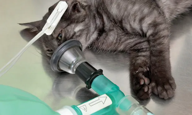
Although less commonly used as an induction agent on account of their dose-dependent cardiovascular effects and the potential for environmental contamination, contemporary inhaled anesthetic agents (eg, isoflurane, sevoflurane) may be used in animals with hepatic disease. Their advantage in this circumstance is the minimal need for hepatic metabolism, and the disadvantage is the cardiovascular depression associated with the high concentrations needed for induciton of anesthesia. To minimize excitement, the patient should be well sedated prior to induction. To minimize the risk for aspiration, inhaled anesthetic induction should be limited to patients with no recent history of vomiting or regurgitation.
Inhaled anesthetics are typically used to maintain anesthesia, but supplementation with cardiovascularly safe medications that reduce the dose requirement is suggested in compromised patients. Short-acting opioids (eg, fentanyl) or opioids that do not require extensive hepatic clearance (eg, remifentanil) are typically recommended for administration by continuous infusion in this circumstance.
ALP = alkaline phosphatase, ALT = alanine aminotransferase, ATP = adenosine triphosphate, AST = aspartate aminotransferase, GGT = gamma–glutamyl transpeptidase