Surgical Treatment for Mammary Tumors
Nicole Ehrhart, VMD, MS, Colorado State University
Mammary tumors are common in dogs and cats. Surgical techniques for mammary tumor removal include lumpectomy, mammectomy, regional mastectomy, unilateral mastectomy, bilateral mastectomy, and radical mastectomy. The ideal technique for each case depends on the species and the number, size, and location of mammary tumors.
In all procedures, the surgical wound should be closed in 2 to 3 layers (deep SC tissue, SC tissue, skin). Consultation with an oncologist for follow-up chemotherapy is recommended when a malignant tumor is diagnosed.
Mammary Tumors in Dogs
In dogs, 50% of mammary tumors are benign. History of a benign mammary tumor does not indicate that subsequent tumors will also be benign. Dogs with a history of benign mammary tumors have a higher chance of developing malignant masses.
Approximately 50% of malignant mammary masses in dogs will metastasize. Therefore, all suspected or confirmed mammary tumors should be thoroughly staged for evidence of metastatic disease. Following physical examination, staging tests should include CBC, serum biochemistry profile, urinalysis, 3-view chest radiography for metastasis check, evaluation of peripheral lymph nodes, and abdominal ultrasonography. Although cytology is not useful for distinguishing benign from malignant tumors, it will help identify other tumor types (eg, mast cell tumor) that can occur in the same location.
Mammary tumors in dogs should be removed by the simplest method (Table) that removes all disease with clean margins. Often, combinations of techniques are used to address disease on opposite mammary chains and when multiple tumors exist.
Table: Recommended Techniques for Mammary Tumor Removal
Once the patient is anesthetized and the surgical area is clipped, it is common to find additional mammary tumors that were not readily palpable with the dog awake. Pet owners must be informed that a revised surgical plan may be required if additional mammary masses are discovered. It is important to obtain owner permission for additional surgery.
Mammary Tumors in Cats
In cats, more than 80% of mammary tumors are malignant, and a high percentage of these masses metastasize. Thorough staging (eg, 3-view thoracic radiographs and abdominal ultrasound) following baseline examination and laboratory tests is essential to determine disease stage.
Because feline mammary tumors are so often malignant and highly metastatic, many cats that develop mammary tumors rapidly develop additional tumors. All mammary tissue should therefore be removed in cats with mammary tumors. This is typically done with unilateral mastectomy followed by removal of the remaining contralateral mammary chain 2 to 4 weeks later. The staged mastectomy allows the skin to stretch between procedures, preventing the respiratory difficulty and excessive pain associated with bilateral mastectomy in a single surgical procedure. As in dogs, the surgical wound should be closed in 2 to 3 layers.
Submitting Resected Tissue
All resected tissue should be submitted for histopathologic analysis. Multiple masses should be submitted in separate containers and clearly labeled. Both malignant and benign masses may be present in the same patient.
What You Will Need
Standard surgical pack
Electrocautery equipment
Absorbable monofilament suture
Step-by-Step: Surgical Treatment for Mammary Tumors
Procedure 1: Lumpectomy
Lumpectomy is used to remove small, BB-pellet–sized (<5 mm in diameter), freely movable masses that are not located directly under a nipple. Remove a small cuff of normal tissue around the mass to ensure all tumor cells are extracted.
Procedure 2: Mammectomy
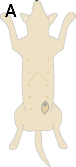
Mammectomy is the removal of a single mammary gland, including the nipple and skin overlying the gland (Figure A). Mammectomy is a good choice when the mammary mass is directly under the nipple or is fixed to the overlying skin. It should not be used for masses fixed to the underlying rectus fascia.
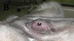
The mammary tissue (M) is superficial to the ventral fascia of the rectus muscle and typically has a layer of fat directly under the glands (Figure B).
Remove all tissue down to the rectus fascia to ensure removal of the mammary gland. Do not remove the rectus fascia or muscle.
Procedure 3: Regional Mastectomy
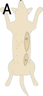
Regional mastectomy is indicated when a mammary mass is located between 2 glands or when multiple small tumors are present in a section of the mammary chain (Figure A). This technique should not be used for masses fixed to the underlying rectus fascia.
Remove the skin, mammary tissue, and underlying fat (Figure B). Do not remove the rectus fascia or muscle.
As necessary, dissect the superficial inguinal lymph node and submit the sample separately for histologic evaluation to further determine the disease stage.
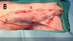
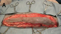
Procedure 4: Mastectomy
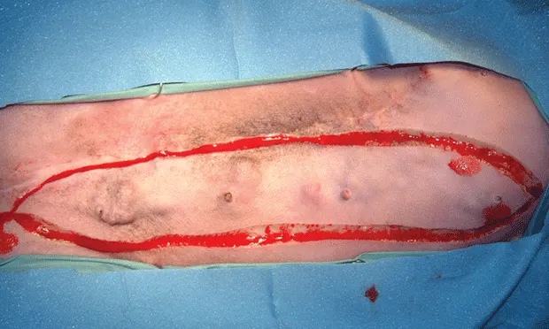
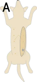
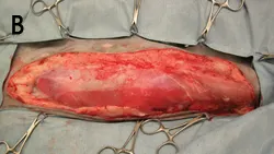
Unilateral mastectomy involves removal of all mammary tissue on one side of midline (Figure A). The mammary tissue extends from midline to the nipple line plus an equal distance lateral to the nipple line.
Make an elliptical incision from the most cranial nipple to the most caudal nipple so that the medial extent of the ellipse is at midline and the lateral extent of the incision is equidistant on the lateral side of the nipple line (Figure B).
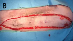
Remove all skin, mammary tissue, and fat down to the rectus fascia. Do not remove the rectus fascia or its underlying muscle.
During caudal dissection, ligate the caudal superficial epigastric artery and vein to prevent excessive bleeding. These vessels run cranially under the mammary glands medial to the inguinal ring and anastomose with the cranial superficial epigastric vessels.
The superficial inguinal lymph node is typically contained within the fat underlying the inguinal mammary tissue. If possible, dissect this node and submit it separately for histologic evaluation to help further determine disease stage.
Unilateral mastectomy can be combined with lumpectomy on the opposite chain. If both mammary chains require mastectomy because of multiple masses, remove the side with the most disease first. Then remove the second mammary chain 2–4 weeks later. This will result in minimal complications related to skin tension.
Author Insight
Bilateral mastectomy performed as a single procedure can often result in unacceptable skin tension and possible respiratory compromise (especially in cats), and has a high complication rate. Staged unilateral mastectomy is recommended when disease extent requires removal of both mammary chains.
Procedure 5: Radical Mastectomy
Radical mastectomy involves removal of the mammary tissue and underlying rectus fascia, muscle, or body wall (Figures A and B). It is indicated for mammary tumors fixed to the underlying body wall. In general, removal of fascia or partial-thickness body wall does not require body wall reconstruction. If full-thickness body wall resection is required, reconstruction methods need to be followed to prevent herniation of abdominal contents.
In cats, the mammary tissue extends to skin overlying the caudal thorax. It is difficult to avoid penetrating the thoracic cavity in cats when partial-thickness body wall resection is attempted during radical mastectomy of the cranial mammary glands. Be prepared to ventilate during anesthesia should inadvertent penetration of the thoracic cavity occur.