Small Animal Parasites That You Want to Forget (But Should Not)
Andrew R. Moorhead, DVM, MS, PhD, DACVM (Parasitology), University of Georgia
Remember learning about the common small animal parasites: heartworm, roundworm, flea tapeworm, Giardia spp, to name a few? In addition, other important parasites that are not as common were taught but maybe not as emphasized. Although the following parasites may not be diagnosed frequently, they are encountered often enough that they should not be forgotten.
Physaloptera spp
The genus Physaloptera, also known as the stomach worm, has sporadic distribution and prevalence and, as its common name implies, lives in the stomach of the definitive host. Transmission occurs by ingestion of an arthropod intermediate host or mammalian paratenic host.1 The diameter of Physaloptera spp worms is the same as that of Toxocara spp but lengthwise the worms are shorter.1 When attached to gastric mucosa in the host, this collar can cause local gastric irritation and bleeding.1 Acute or chronic vomiting also can occur secondary to gastric irritation. In addition, the worms reportedly can cause anorexia, melena, and anemia, secondary to any hemorrhaging.2
Diagnosis is normally made when a worm (usually an adult) is found in the pet’s vomitus.3 While not ideal, due to the need for anesthesia, diagnosis is sometimes made on endoscopy.3 Diagnosis by fecal flotation is possible, but the eggs can be difficult to detect, as they typically are too heavy to float in solution. Thus, the fecal sedimentation technique is the preferred means of diagnosis.4
The following treatment regimens have been used, although other protocols5 also have been described:
Dogs — fenbendazole at 50 mg/kg PO once daily for 3 days6
Cats — 2 doses of pyrantel at 5 mg/kg PO 3 weeks apart7 or a single dose of ivermectin at 0.2 mg/kg SC2
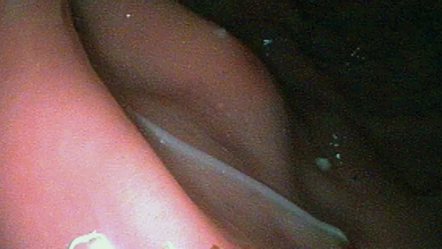
Endoscopic view of Physaloptera spp nematode present in the gastric body. Courtesy of Colin F. Burrows, BVetMed, PhD, Hon FRCVS, DACVIM, University of Florida
Capillarids
The Capillarid class of parasites resemble whipworm eggs. The following capillarids reside in the urinary or respiratory tract of the definitive host.
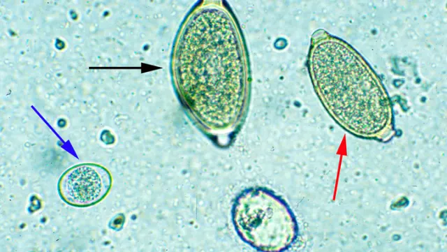
Comparison of a Trichuris spp egg (black arrow) with a capillarid egg (red arrow) and an Isospora spp oocyst (blue arrow). Courtesy of Anne M. Zajac, DVM, PhD, Virginia-Maryland Regional College of Veterinary Medicine
Pearsonema spp—The Urinary Capillarids
Pearsonema plica infects dogs and wild canids,8-10 whereas P feliscati seems to be specific to cats.11-13 Adults embed in the urinary bladder mucosa and shed unembryonated eggs.
The life cycle most likely involves an earthworm paratenic/intermediate host.5 Infection is usually subclinical, although cystitis can occur from a large number of worms being embedded in the bladder mucosa. Infected animals present with hematuria and attempt to urinate frequently.5,14 Observing eggs on examination of urine sediment is definitively diagnostic.14,15 In dogs, successful treatment has been reported after a single SC injection of ivermectin at 0.2 mg/kg16; fenbendazole at 25 mg/kg PO twice a day for 10 days reportedly alleviated signs in an infected cat.17
Eucoleus spp—The Respiratory Capillarids
Adult Eucoleus spp worms are embedded in the nasal or respiratory mucosa and, depending on the specific parasitic species, can be found in both dogs and cats.5
Eucloeus aerophilus is found in foxes, dogs, and cats.18,19 Adult nematodes live in the mucosa of the trachea, bronchi, and bronchioles. Eggs are produced by the worm, coughed up in the sputum, swallowed, and excreted in feces. Infection results after ingestion of infective eggs or potentially an earthworm paratenic/intermediate host. Larvae hatch in the small intestine and then migrate through the bloodstream to the lungs, where they penetrate the alveoli and migrate into the air passages. The prepatent time is suspected to be 3 to 5 weeks. As with infection by the urinary capillarids, respiratory E aerophilus infection is normally subclinical. An occasional to chronic cough and wheezing may be noted. In severe cases, the animal may present with tracheobronchitis, dyspnea, and pneumonia. 20 Definitive diagnosis is made by detecting eggs in fecal flotation, sputum, or transtracheal wash.4 Eggs are greenish brown and rather asymmetric, with a granular shell, bipolar plugs, and single-cell embryo. A single treatment of topical moxidectin at 1 mg/kg and imidacloprid at 10 mg/kg has been successfully used to treat infection in cats.21 Ivermectin, administered once at a dose of 200 µg/kg PO, has been successful in dogs.5
E boehmi is the nasal capillarid of dogs.22 Adults live in the mucosa of the nasal turbinates and sinuses. The life cycle is unknown but as with E aerophilus, the earthworm may be involved. Eggs are passed in feces or in excess nasal mucus. Infection is usually subclinical. The presence of worms and eggs can result in inflammation, which can subsequently cause sneezing and mucopurulent nasal discharge. Definitive diagnosis is made by observing eggs on a smear of nasal discharge or nasal washings (though the eggs can be difficult to recover by nasal swab) or by fecal flotation.23,24 The eggs are symmetric and golden with bipolar plugs, with the egg surface pitted and having a thimble-like appearance. Fenbendazole given at a dose of 50 mg/kg PO once a day for 2 weeks, in conjunction with prevention of coprophagia, results in a resolution of signs in dogs.25 As with E aerophilus, a single oral dose of ivermectin at 200 µg/kg 26 has been used for treatment in dogs as well.
Aelurostrongylus abstrusus—The Feline Lungworm
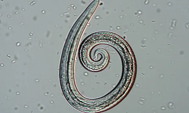
In the southeastern United States, Aelurostrongylus abstrusus should be a differential diagnosis in all coughing cats. Adult worms live in the terminal bronchioles and alveolar ducts. Eggs are deposited in clusters in the lung. The larvae inside the eggs hatch and migrate up the trachea, after which they are swallowed and enter the gastrointestinal (GI) tract. First-stage larvae (L1) are shed in feces.27,28 A snail or slug becomes infected with L1, which then develop into infectious third-stage larvae.29 The slug can then be consumed by a paratenic host. When a slug or the paratenic host is ingested by a cat (ie, the definitive host), the larvae migrate from the GI tract to the lungs. In 5 to 6 weeks, the worms develop into adults, thus completing the life cycle.5,23
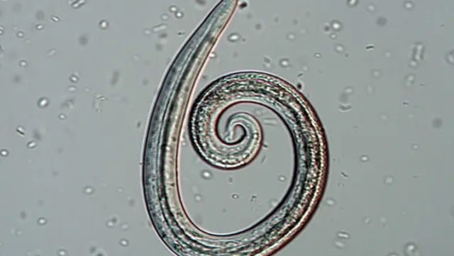
Aelurostrongylus spp L1 larva identified from Baermann fecal sample. Courtesy of Dwight D. Bowman, MS, PhD, DACVM (Hon), Cornell University
Radiographic changes may be evident30,31; however, definitive diagnosis can be based on the results of a Baermann fecal technique for nematode larval identification, which is the most reliable diagnostic method. Alternatively, a zinc sulfate centrifugal flotation can be conducted. A abstrusus larvae have an S-shape kinked tail and subterminal dorsal spine.4 Treatment options include, but are not limited to, the following:
Single topical applications of selamectin at 6 mg/kg (successful in 1 of 3 cats)33
Fenbendazole at 20 mg/kg PO once a day for 5 days or 50 mg/kg PO once a day for 15 days33,34
Single dose of ivermectin at 400 µg/kg SC35
Single topical application of moxidectin at 1 mg/kg, along with imidacloprid at 10 mg/kg36
Single topical application of emodepside at 3 mg/kg, along with praziquantel 8.6%.37
Prednisone may also be used for symptomatic treatment.
Closing Thoughts
Though these parasites are not as common as heartworms, roundworms, or flea tapeworms, they are diagnosed frequently enough to warrant remembering, especially when encountering associated signs in patients. Appropriate diagnostics are essential to identify the correct treatment.