Rehabilitation Therapy: Sciatic Nerve Injury
Jennifer Au, DVM, DACVS, DACVSMR, CCRT, Charleston Veterinary Referral Center, Charleston, South Carolina
Signalment & History
Ms. Red, a 2-year-old, 22.3 kg, spayed hound mix, was presented for evaluation of non–weight-bearing lameness of the left hind limb of 3 weeks’ duration. The dog was healthy, with no known clinical abnormalities prior to ovariohysterectomy (OHE) 3 weeks earlier. While the dog was anesthetized for the OHE, she received an IM penicillin G injection of unknown volume and concentration in the left pelvic limb. On recovery, the dog exhibited a non-weight-bearing lameness on that limb. There had been no change in limb use since that time.
Previous & Current Treatments
Unknown doses of oral amoxicillin and dexamethasone had been administered postoperatively. No change in use of the limb was noted following administration. Two weeks after the OHE, the dog was evaluated by a second veterinarian and placed on carprofen at 75 mg (3.4 mg/kg) PO once daily. The dog was presented for evaluation three weeks after the OHE, with no change in limb use.
Physical Examination
Physical examination showed non–weight-bearing lameness of the left hindlimb, with muscle atrophy primarily associated with the distal one-half to one-third of the caudolateral muscles of the thigh (biceps femoris, semimembranosus, semitendinosus) and the craniolateral tibia. When standing, the dog would occasionally lower the left hindfoot to the ground, resting in a knuckled position on the dorsal aspect of the digits. Superficial abrasions to the dorsal aspect of the left hind digits were present. When the left hindfoot was manually placed in the appropriate position, the dog was able to stand on her left side with minimal support while the right hindlimb was slowly lifted. The orthopedic examination was unremarkable.
Notable findings on neurologic examination included absent conscious proprioception of the left hindlimb, normal reflexes (patellar, cranial tibial, gastrocnemius), and decreased flexion at the hock on the withdrawal reflex. Normal range of motion was present at the coxofemoral, stifle, and tarsal joints. Superficial sensation to the sciatic dermatomes appeared intact when the skin was pinched with hemostats.
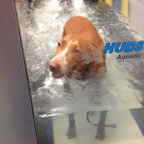
Initial presentation for rehabilitation therapy: Ms. Red walking on dry land.
Diagnosis
Definitive
Left peripheral sciatic nerve injury, primarily of the peroneal/fibular branch
Differentials
Neurapraxia (temporary disruption of function) versus axonotmesis (disruption of axons without disruption of the surrounding connective tissue of the nerve)1
Rule out for nerve injury occurring secondary to IM injection (penicillin G) at time of OHE versus other causes of nerve injury2-4
Therapeutic Recommendations
In-house rehabilitation
8 sessions, 2 times a week, with reevaluation on completion of the 8 sessions
Home exercise plan
Provided to owner after initial session
Continue carprofen as previously directed by referring DVM
Therapeutic Modalities
Exercises emphasizing proprioception, balance, and weight-bearing
Therapeutic laser
Underwater treadmill
Land/terrestrial treadmill
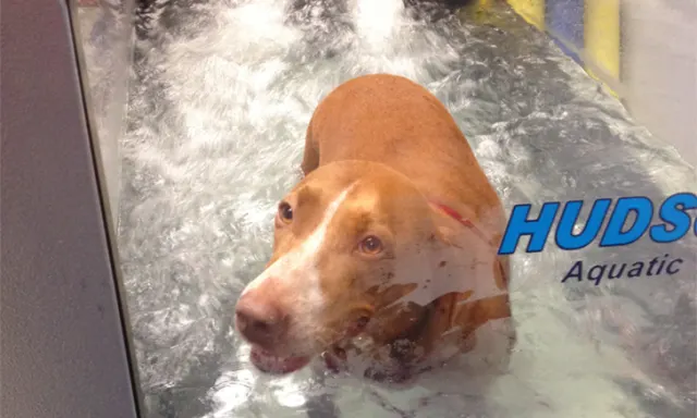
Sixth rehabilitation therapy session: Ms. Red walking in underwater treadmill.
Rehabilitation Goals
Short-term (days/weeks)
Prevent progressive muscle atrophy
Encourage weight bearing and limb use
Aid proprioceptive function
Long-term (weeks/months)
Return to normal muscle mass
Return to normal limb use when walking or trotting
Minimize or eliminate proprioceptive deficits during walking and trotting
Outcome Measurements
Subjective
Limb use and placement
Neurologic examination
Objective
Gulick spring tension tape measurement of the thigh muscle circumference (Table 1)
Measurements as taken one-third the distance from the greater trochanter to lateral fabella
Thigh Muscle Circumference Using a Gulick Tape Measure
Therapeutic Exercises
A variety of active exercises were used throughout the dog’s therapy. Initially, exercises were selected to encourage the patient to toe-touch, using appropriate placement of the paw, by focusing on disruption of balance (ie, perturbation); with the dog standing, proprioception was challenged through perturbation or weight-shifting motions. This exercise is designed to secondarily result in placement of the affected limb.
As the dog’s condition improved, the forelimbs were positioned on an aerobic step that allowed weight-shifting to the hindlimbs and gradual increase in the level of difficulty. To challenge the dog further, she was placed on a variety of unstable surfaces (eg, physioball, balance board, wobble disk). As proprioception and placement of the affected limb improved, exercises to encourage weight bearing and functional limb use were added, including 3-legged stand, walking over an unstable surface (air mattress, foam mat, dog bed), and walking over obstacles.
Modalities
A series of modalities or tools, including therapeutic laser, underwater treadmill (UWTM), and land/terrestrial treadmill, were also used. Therapeutic laser and UWTM were used during each session. Land/terrestrial treadmill walking was added during the third session.
A therapeutic class 4 laser (808/905 nm, 4J/cm2; MLS Therapy Laser, celasers.com) was used along the course of the sciatic nerve, from the sciatic notch to the level of the proximal tibia. This low-level laser therapy (LLLT; also called cold laser) followed the course of the sciatic nerve and the peroneal/fibular branch as it innervated the cranial tibial muscle, the primary flexor of the tarsus. Studies in rats have indicated that laser therapy can help with nerve recovery and appears to enhance production of proteins and growth factors that encourage axonal sprouting and accelerate healing.<sup5 sup>
UWTM therapy was introduced to encourage limb use, gain muscle mass, and stimulate superficial sensory stimuli of the affected limb. The water level was set even with the greater trochanter and adjusted as needed based on the dog’s limb use, thereby allowing a normal gait pattern of the three unaffected limbs without the dog attempting to swim. A custom-made dorsal flexion device was applied to the affected limb from the hock to the digits. This device aided in dorsal flexion of the hock and extension of the digits to promote appropriate paw placement in the water. During the first UWTM interval, the dog used the affected limb approximately 20% of the time, showing frequent knuckling. However, by the third interval, she was using the limb 40% to 50% of the time, although still with frequent knuckling. The dog’s use of limb progressively improved; by the eighth and final session, approximately 2.5 months after the initial injury, she was using the limb 100% of the time with no evidence of knuckling while on the UWTM.
After the dog ambulated during UWTM therapy (ie, at end of each session), massaging jets were used to facilitate muscle relaxation and phasic stimulation of sensory receptors and to decrease nociceptor hypersensitivity. During the third session, inclined walking on a land/terrestrial treadmill was added to help encourage proprioceptive training and limb use and rebuild muscle mass. The patient responded better to walking on the UWTM than on the land/terrestrial treadmill. The UWTM was used as a primary means of treadmill therapy with each session, whereas, the land/terrestrial treadmill was attempted but not aggressively pursued because of the dog’s limited response to this therapy.
Home Exercises
The owner was instructed in completion of a series of exercises to be performed at home. Initially, these exercises consisted of proprioceptive and weight-bearing activities. For example, one exercise consisted of placing the dog’s front paws on a slightly elevated surface (eg, stairs, footstool, couch cushion) and gently rubbing or rocking the dog from side-to-side or front-to-back to encourage placing weight on the affected limb. Later in the recovery process, tug-of-war, obstacle courses, and challenged walking (uphill, sideways, backward) were added.
At completion of rehabilitation therapy: Ms. Red walking on dry land.
Reevaluation
The dog was reevaluated after a series of 8 rehabilitation sessions performed over a 7-week period. Mild-to-moderate weight-bearing lameness of the left hindlimb was evident, but the dog would use the limb consistently at a walk. At a trot, she intermittently hopped or held the limb up for 1 or 2 steps. Muscle atrophy was still present as previously described but had improved both subjectively and on follow-up measurement of the thigh circumference (Table 1). Decreased withdrawal at the hock remained but demonstrated improvement on subjective evaluation.
An additional series of 8 rehabilitation sessions was recommended; however, the owner elected to discontinue therapy at this point. The owner was encouraged to continue the home exercises and present the dog for a recheck follow-up if improvement stalled or worsened. Approximately 6 weeks after discharge from the rehabilitation program, the owner sent video confirmation of the dog using the affected limb consistently while walking, running, and playing with other dogs.
Discussion
Injury to the sciatic nerve is an uncommon but potential complication of IM injections into the caudal thigh musculature, partially because of the size of the nerve and its relatively peripheral location. Injections within (intraneural, intrafascicular) or around (perineuronal) a nerve can result in injury.2 The drug itself, carrier vehicle, nerve involved, needle type, location of the injection, and pressure of the injection can all influence the onset of nerve injury.2-4 Side effects associated with injection can range from pain at the injection site to necrosis of the nerve.3
Two months following rehabilitation therapy: Ms. Red playing with another dog.
The 3 types of possible nerve injury are:1
Neurapraxia, resulting in temporary loss of motor and sensory function
Axonotmesis, resulting in disruption of the axon and myelin sheath but not the surrounding connective tissue (epineurium and perineurium), allowing a guided route for axon regrowth
Neurotmesis, resulting in complete loss of continuity of the axon and surrounding connective tissue1
Recovery from neurapraxia can take hours to months, whereas axonotmesis requires months for recovery.1 In cases of neurotmesis, recovery of function is not possible without surgical intervention. In this case, neurotmesis was ruled out based on the intact sciatic reflexes and intact superficial pain sensation. However, neurapraxia or axonotmesis with partial injury was possible based on the neurologic examination. Electromyography (EMG) and nerve conduction studies can be performed to further evaluate the degree of nerve injury and prediction of recovery.1,6,7
The initial and final evaluations were performed by the same veterinarian, and all therapy sessions were performed by a veterinarian or licensed veterinary technician certified in canine rehabilitation (CCRP, CCRT). Sessions lasted approximately 1 to 1.5 hours. After the initial evaluation, therapy was recommended twice weekly for 8 sessions, with reevaluation to assess the progress at completion of the 8 sessions. Thigh muscle circumference was measured as an objective assessment of response to therapy. Joint range of motion was deemed normal on the initial evaluation and, therefore, was not recorded on initial or follow-up assessments. Other, more objective assessments of response to treatment were not available.
Active exercises involve those the patient could undertake, under the direction of the rehabilitation therapist or owner, to facilitate use of specific muscles, joints, or nerves in combination. Passive exercises are those performed by the rehabilitation therapist or owner with little to no active participation by the patient. Because active exercises engage more muscle groups and nerves than passive exercises do, they are typically preferred over passive exercises if the animal is able to perform them. Active exercises can be used to engage the patient’s proprioceptive system and encourage limb placement; proprioception is challenged through perturbation or weight-shifting motions. When an animal’s balance is challenged, the normal response is to prevent falling by engaging a series of nerves and muscles. This often results in placement of the affected limb. Once the animal is placing the limb, exercises to encourage limb use can be added to the treatment protocol.
This case report demonstrated a rehabilitation program involving a series of active exercises and treatment modalities to aid in limb function after onset of peripheral neuropathy secondary to an IM injection.