Feline Injection-Site Sarcoma Assessment
Timothy M. Fan, DVM, PhD, DACVIM (Oncology, Internal Medicine), University of Illinois at Urbana–Champaign
In the Literature
Ferrari R, Di Giancamillo M, Stefanello D, et al. Clinical and computed tomography tumour dimension assessments for planning wide excision of injection site sarcomas in cats: how strong is the agreement? Vet Comp Oncol. 2017;15(2):374-382.
The Research ...
Cats appear to be uniquely vulnerable to sarcoma development as a consequence of localized irritation and inflammation following the injection of vaccines and other medical therapeutics1-3; these tumors are referred to collectively as injection-site sarcomas (ISSs). Biologically, ISSs are locally invasive and grow rapidly. The most effective treatment outcomes rely on complete gross surgical resection of macroscopic tumor burdens.4-6 As such, assessment methods for accurately defining tumor margins are of paramount importance for guiding appropriate surgical planning and maximizing long-term cure rates in affected cats.
This prospective study examined the agreement between the measurement (length and width) of ISS sizes when assessed by digital calipers (clinical method) and contrast-enhanced CT (Figure). Over a span of 11 years at a single institute, the tumor dimensions of 53 pet cats with ISSs were evaluated before surgery by a single surgeon using the clinical method and by a single radiologist using the CT method.
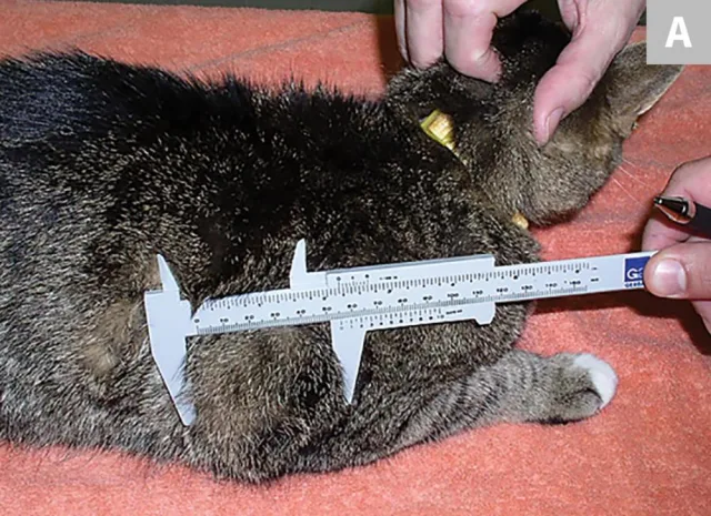
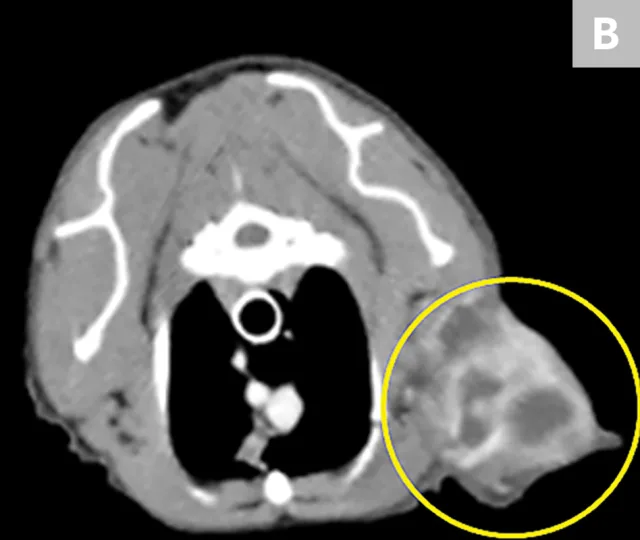
Clinical measurement with manual calipers (A) of an ISS involving the soft tissues overlying the right distal lateral humeral region of a cat. A transverse view with contrast-enhancing CT (B) in the same patient readily identifies a multilobulated soft tissue tumor (yellow circle) with discrete regions of peripherally enhanced vascular growth and proliferative tumor tissues.
Caliper and CT methods tended to be in agreement directionally; however, CT methods consistently estimated greater tumor sizes (length and width) as compared with the clinical method. The agreement between clinical and CT methods became more divergent as a function of increasing tumor size. In addition, the characterization of ISSs as adopting irregular-shaped growth patterns (vs spheroid or ovoid) was identified more readily by CT methods.
Integration of the findings derived from both clinical and CT methods for ISS margin assessment should be considered the best clinical practice in planning the successful surgical resection of ISSs in cats.
...The Takeaways
Key pearls to put into practice:
Both digital caliper (clinical) and contrast-enhanced CT methods for assessment of ISS tumor dimensions tend to be in agreement and should be used in combination to maximize the success of ISS resection in cats.
Contrast-enhancing CT methods have the advantage of identifying regional physiologic processes, including inflammation and vascularization, which cannot be readily appreciated by caliper methods.
Irregular growth patterns (extending neoplastic tendrils) displayed by ISSs can be identified more easily via contrast-enhancing CT methods.
Prospective studies are required to definitively determine if a singular method (clinical vs CT) is more predictive of achieving long-term surgical cures.