Determining Whether a Dog Is Spayed
Milan Hess, DVM, MS, DACT, Colorado Veterinary Specialists & Animal ER , Littleton, Colorado

History
Queenie, a mature adult female boxer, was evaluated following adoption from a local shelter. The shelter presumed she had been spayed because of a scar on her ventral midline consistent with an ovariohysterectomy (OHE); however, within a month of adoption, Queenie developed vulvar swelling and began to attract male dogs.
Queenie, a mature female boxer, was presented after developing vulvar swelling and attracting male dogs.
Examination
Queenie was bright and alert; had a body condition score of 5/9; and showed minor mammary gland development, moderate vulvar swelling, and slight serosanguinous vaginal discharge. The remainder of the examination was unremarkable.
Related Article: Ovarian Remnant
Initial Diagnostic Evaluation
A vaginal cytology sample was collected to determine if the vulvar swelling and discharge were related to elevated estrogen levels. The cytology sample swab was rolled on the slide, air dried, and stained with Diff-Quik before being examined. The sample contained predominately large, cornified superficial cells with pyknotic nuclei and few other cells or background debris (Figure 1).
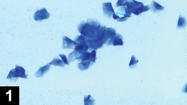
Vaginal cytology with large cornified epithelial cells or superficial cells indicating elevated estrogen concentrations.
The presence of cornified epithelial cells confirmed estrogen stimulation. To determine if the estrogen exposure was endogenous or exogenous, the owner was thoroughly questioned about recent administration of oral or injectable estrogens and possible contact with human transdermal hormone replacement therapy (HRT). Dogs are very sensitive to HRT and can show signs of estrogen stimulation after contact with persons, clothing, or bedding contaminated with HRT.1 Additional testing to confirm the presence of ovarian tissue was recommended.
ASK YOURSELF
What additional diagnostic tests can be performed to determine if this patient has ovarian tissue?
A. Luteinizing hormone test
B. Serum progesterone concentration
C. Anti-Müllerian hormone assay
D. Ultrasound
E. All of the above
A combination of history, examination findings, and diagnostic tests are used to diagnose the presence of ovarian tissue. The same strategy can be used to determine if a bitch is intact, spayed, or has an ovarian remnant (OR).
Luteinizing hormone (LH) is negative (<1 ng/mL) in intact bitches or those with ovarian remnant syndrome (ORS). In addition, LH is negative in bitches exposed to endogenous or exogenous estrogen.2 LH tests are not performed if the patient is currently showing signs of estrogen stimulation. A negative result in a nonestrogenized bitch is consistent with the presence of ovarian tissue.
Serum LH is positive (>1 ng/mL) in bitches without ovarian tissue unless the test has detected the LH surge for that cycle. The LH test can be used to screen for ovarian tissue if the bitch does not show evidence of estrogen stimulation at the time of evaluation. Two positive tests performed several days apart (to ensure the sample was not taken during the LH surge) provide conclusive evidence that ovarian tissue is not present. Major veterinary laboratories perform LH tests, and an LH test kit is also available (Witness LH, zoetis.com) for in-house use.
Progesterone concentrations are consistently <0.2 ng/mL in bitches without ovarian tissue and rise following ovulation in intact bitches or those with ORS. Documenting serum progesterone >5 ng/mL in a bitch with evidence of estrogen stimulation 3–4 weeks prior confirms the presence of ovarian tissue. In addition, progesterone concentrations <1 ng/mL alone do not rule out ORS, as the dog may be in between heat cycles (anestrus). Quantitative progesterone measurements are performed at major veterinary laboratories.
Tips for Successful ORS Surgery
Make a large incision to allow complete visualization of the abdomen.
Thoroughly examine all mesenteric surfaces.
Submit the excised tissue for histopathological evaluation.
Perform the surgery 2–4 weeks after the bitch goes out of heat.
This is when the luteal tissue (progesterone-producing tissue) is prominent and the remnant is most easily located.
Anti-Müllerian hormone (AMH) is produced solely by the ovaries in females,3 so AMH measurement can be used to distinguish between intact and spayed bitches. The AMH ELISA can be used successfully to diagnose ORS in bitches. The in-house lateral flow test is not recommended for ORS screening as it is not sensitive enough to detect some ORS cases.2 AMH is not suppressed by estrogen and can be used at any time in post-pubertal bitches to evaluate for the presence of ovaries. The AMH ELISA is currently performed at the Animal Health Diagnostic Center at Cornell University; the Clinical Endocrinology Laboratory at University of California, Davis; and AViD Laboratories.
Although ovarian tissue can be seen on ultrasound, it is most successfully employed when the bitch is in estrus and has active follicular tissue and the ovaries are in their normal location. Ovarian remnants can be located anywhere in the abdomen and can be challenging for the most experienced ultrasonographer to find, particularly if the tissue is inactive at the time of evaluation or in an unexpected location.
ORS is caused by incomplete or improper removal of the ovaries during OHE or ovariectomy (OVE).4 Ectopic ovarian tissue does not occur in the bitch. Surgeon experience is not correlated with ORS incidence. The right ovary is affected most frequently, and the interval from OHE/OVE to diagnosis is 1 month–10 years.3 Factors thought to increase risk for ORS include inadequate incision length, poor exposure, and failure to examine the ovaries after removal.3
Treatment
Exploratory laparatomy and excision of OR(s) is the treatment of choice. The majority of remnants are found in the region of the pedicles. A complete exploratory should be performed to ensure all ovarian tissue is removed. Ovarian remnants can also be created by fracturing of the ovary during OHE/OVE and subsequent revascularization of the ovarian tissue. These remnants can be located anywhere in the abdomen and can require a lengthy search to uncover.
The Take-Home
A combination of history, physical examination findings, and hormone testing is used to determine if a bitch is intact or has an ovarian remnant.
Hormone testing choice and timing depends on presence of current estrogen stimulation and where the bitch may be in her cycle
HRT = hormone replacement therapy, OHE = ovariohysterectomy