Cough, Diarrhea, & Incorrect Assumptions
History (Day 0)
Carter, an intact male Yorkshire terrier puppy (14 weeks of age), was purchased in the Canadian province of Saskatchewan and shipped as live air cargo to the owner on Prince Edward Island. The puppy was presented for a cough of approximately 3 days duration. Vaccinations were up-to-date. The puppy had been purchased from a breeder about 4 weeks before presentation.
Examination
Carter appeared bright and alert. Temperature and pulse were normal. Body condition score was normal. Increased bronchovesicular sounds were detected on auscultation. The puppy coughed during examination, and both ocular and nasal discharges were noted. Kennel cough, collapsing trachea, and lungworm infection were considered likely as possible rule-outs. Distemper and pneumonia were not considered, as the breeder had an excellent reputation and vaccinations were up-to-date.
Treatment
Atlantic Canada has a high incidence of fox lungworm (Crenosoma vulpis) infection —acquired by ingestion of gastropod intermediate hosts and resulting in a nonfatal, chronic cough—in dogs.1 Diagnosis occurs by detection of first-stage larvae via Baermann fecal examination. Because fecal larval shedding tends to be intermittent, false-negative Baermann results can occur, and it is common in this region to deworm dogs presenting with signs of cough and pursue diagnostic testing only if the condition does not resolve.
Related Article: Differentiation of Parasites & Pseudoparasites
Carter was treated with a 3-day course of fenbendazole (50 mg/kg PO q24h). The cough greatly improved but did not resolve completely.
History (Day 8)
Within 5 days of receiving the last deworming treatment, Carter developed diarrhea.
Examination
The results of the physical examination were unchanged from the previous examination.
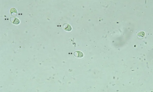
Figure 1. Giardia duodenalis cysts detected on zinc sulfate centrifugal fecal flotation. Note the artifact in the cysts, which appears as a vacuole or empty space, caused by the osmotic damage from the high specific gravity of the flotation media.
Diagnostics
Radiographs showed a slight increase in density in the lung field. A fecal sample was submitted, and a zinc sulfate centrifugal fecal flotation examination was conducted. A large number of Giardia duodenalis cysts were detected on the fecal flotation examination (Figure 1). CBC and serum chemistry panel were not performed at this time.
Treatment
An additional 5-day course of fenbendazole (50 mg/kg PO q24h) was prescribed to treat giardiasis.
History (Day 20)
About 1 week after the second course of fenbendazole was completed, the cough returned and diarrhea became markedly worse, containing streaks of frank blood and mucus.
Examination
Carter was listless, was anorexic, and had abdominal pain on palpation.
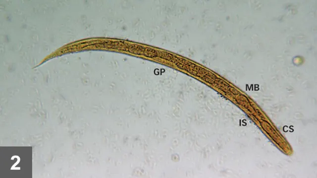
Figure 2. Strongyloides stercoralis first-stage larvae (L1) recovered by Baermann fecal examination (iodine stained and killed). Note the distinctive esophageal morphology: muscular corpus (CS) followed by a narrow isthmus (IS), terminating in a rounded muscular bulb (MB). Note also the prominent genital primordial cell (GP) appearing midbody along the intestine.
Diagnostics
A fresh fecal sample was collected and submitted for both zinc sulfate centrifugal flotation and Baermann examination. The fecal flotation results were negative. The Baermann examination detected first-stage larvae of Strongyloides stercoralis (Figure 2).
Treatment
Ivermectin (200 µg/kg PO, repeated in 1 week) was administered. There was marked clinical improvement within several days of receiving the first treatment, and all signs were resolved by the time the second dose was given. No first-stage larvae were detected on posttreatment Baermann fecal examinations conducted 7 days after the first dose and 7 and 30 days after the second dose of ivermectin.
Diagnosis
Strongyloidiasis
Discussion
When forced to cut corners when resources are limited, it is prudent to make educated guesses when specific information is not available. The assumption that the initial cough condition was probably a result of C vulpis infection was reasonable given the region. Lungworm is the most likely cause of chronic cough in dogs in that region and presumptive treatment is a common practice.1 Assuming that the diarrhea was from giardiasis—a common cause of diarrhea in dogs under 1 year of age—considering the fecal flotation result was also reasonable. Because of the high prevalence of infection in puppies, most of which are subclinical infections,2 the possibility exists that the presence of Giardia spp could be incidental in some cases of canine diarrhea. Both of these assumptions (ie, C vulpis infection as the cause of cough, Giardia spp infection as the cause of the diarrhea) were wrong and resulted in a potentially deadly delay. This case serves as a reminder that cutting corners, even when it appears most reasonable to do so, can result in terrible consequences.
Signs of cough preceding the onset of diarrhea by several days are frequently observed in infected dogs.
Strongyloides stercoralis infects the small intestine of humans, primates, and occasionally dogs.3 Infection is more common in the tropics and subtropics, but S stercoralis can be found throughout the world. Infection is acquired by ingestion of infective third-stage larvae (L3) or through direct skin penetration of the L3. Transmammary transmission occurs in dogs and is an important exposure route in neonates.4 Larval stages undergo either a somatic migration (encysting in various tissues for prolonged periods of time) or a tracheal migration (migrating to liver and/or lungs, coughed up, and swallowed to reside in the small intestine). The respiratory signs in the puppy in this case were caused by tracheal migration of the larval stages. Signs of cough preceding the onset of diarrhea by several days are frequently observed in infected dogs.3
Related Article: Labored Breathing: Hairballs, Parasites, or Worse?
Infection with S stercoralis in humans can be fatal. Individuals surviving the initial infection may suffer fatal infections many years later if they become immunosuppressed from hyperinfection or autoinfection from somatic tissue larvae resuming their development.5,6 Dogs do not appear to be a significant source of infection for humans; however, because of the serious consequences of human strongyloidiasis, prompt diagnosis and treatment of infected dogs is prudent.3
No anthelmintics are currently approved for use in the treatment of dogs infected with S stercoralis. Ivermectin and fenbendazole have been used with some success in treatment of dogs.<sup7,8 sup>
Outcome
Clinical improvement was observed in response to several courses of fenbendazole treatment in this patient; however, the infection persisted requiring further treatment with ivermectin.
GARY A. CONBOY, DVM, PhD, DACVM, is faculty member of Atlantic Veterinary College’s pathology and microbiology department in Charlottetown, Prince Edward Island, Canada. He is a charter diplomate of the American College of Veterinary Microbiology’s board specialty in parasitology and co-author of Veterinary Clinical Parasitology’s seventh and eighth editions. For more than 20 years, his main research focus has been nematode lungworm parasites of wild and domestic canids. He is a University of Minnesota graduate.