Corneal Ulceration in the Guinea Pig
The guinea pig is a popular exotic companion mammal with its own set of care requirements and health issues. However, the author has observed that guinea pigs are hardy animals and can enjoy very good health when provided appropriate hygiene and a proper diet. Their friendly personalities and relatively small size make them a good pet choice for families with children.
Related Article: Environmental Enrichment for Small Mammals
Overview
Despite their popularity as both pets and laboratory research rodents, there is a relative lack of published information on ocular disease in guinea pigs. Anecdotal evidence has suggested that guinea pigs have a high prevalence of ocular lesions.1 A comprehensive study of 1000 guinea pigs from both research and pet populations revealed that high proportion of ocular abnormalities.1
Anecdotal evidence has suggested that guinea pigs have a high prevalence of ocular lesions.1
Guinea pigs have a paurangiotic retina: blood vessels in the retina are visible only near the optic disk, which makes the retina appear avascular on examination.2,3 Their eyelids are open from birth, and they have small third eyelids.2,3 Compared with other mammals, guinea pigs produce very small amounts of tears, which can predispose them to conditions that can affect corneal health (eg, ulcerative keratoconjunctivitis).2-4
Corneal ulceration can be attributed to trauma associated with bedding, hay, caging, a companion, hair impingement on the cornea (trichiasis), decreased tear production, or infection.1,4,5 Because of their naturally low tear production, some breeds may be predisposed to ulcerative keratoconjunctivitis (ulceration and corresponding inflammation of the cornea or conjunctiva).1,2 Another potential cause of corneal lesions in guinea pigs is neoplasia, specifically lymphosarcoma,2,6,7 which reportedly can infiltrate the cornea.1,2,6,7
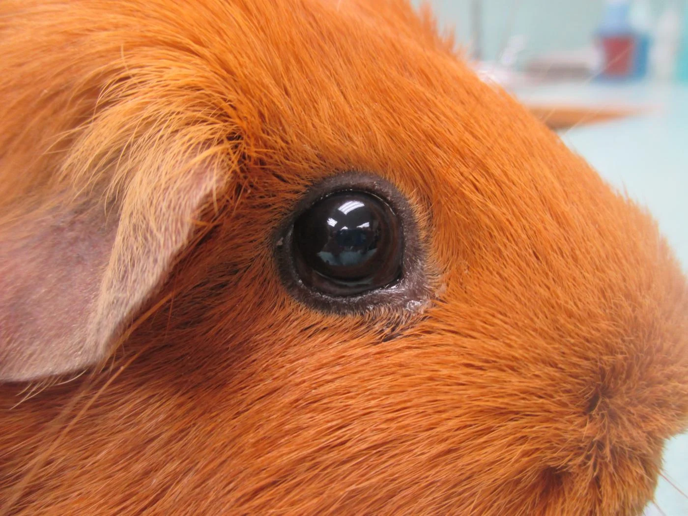
Figure 1.
Short-haired guinea pig with normal eye.
Diagnosis
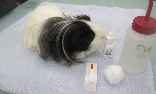
Diagnostic ophthalmic testing is performed similar to that done for other mammals. In guinea pigs, tear production is most accurately measured with the phenol red thread tear test rather than with Schirmer tear test strips<sup.2-4sup> Fluorescein staining can be used to evaluate corneal health and integrity. Diagnosis of corneal ulceration is obtained by direct ophthalmic examination and fluorescein staining.2-4 If corneal ulceration is present, the lesion will be revealed when the affected area of the cornea absorbs the fluorescein dye.
Related Article: Corneal Cytology & Culture Collection
Treatment
Treatment of corneal ulceration in guinea pigs is based on the cause as well as the condition of the individual patient. Any foreign material that is present should be removed either by flushing or gentle debridement. If ulceration is attributed to trichiasis, hair removal or surgical correction of the eyelid can potentially resolve the condition Most guinea pigs tolerate topical eye medications well, either in liquid or ointment form. Ointment medication can be advantageous because it remains on the surface of the cornea for a longer duration, allowing extended treatment intervals. Steroid medication, either systemic or topical, should be used sparingly, as there is potential for adverse effects (eg, recrudescence of nonclinical secondary bacterial or Chlamydophila caviae infection). Steroids are also contraindicated in the presence of a corneal ulcer. The use of cone collars is not recommended in guinea pigs because of their potential reluctance to eat and the challenge associated with keeping the collar in place.
Closing Remarks
When performing a comprehensive physical examination on guinea pigs, be sure to examine their eyes closely and conduct appropriate ophthalmic testing if indicated. If corneal disease is identified, evaluation for environmental, traumatic, or systemic factors that may have contributed to the condition is warranted.
Related Article: Venipuncture in Small Exotic Companion Mammals
Tracey K. Ritzman, DVM, DABVP (Avian, Exotic Companion Mammal), is an associate veterinarian at Cascade Hospital for Animals in Grand Rapids, Michigan. Dr. Ritzman is the only board-certified veterinarian in exotic companion mammal practice in the State of Michigan. She received her DVM from North Carolina State University.