The Case: Gastroenteritis or Something Else?
Gretchen Statz, DVM, DACVECC, Antech Diagnostics, Veterinary Emergency and Specialty Care, Indianapolis, Indiana
Barak Benaryeh, DVM, DABVP, Spicewood Springs Animal Hospital, Austin, Texas
PRESENTATION 1
A 7-year-old castrated male Lhasa apso mix was presented to the emergency clinic at 11:30 pm. The dog had vomited (unwitnessed) multiple times during the day. The owner also reported that the stools may have been dark. The dog had a history of eating organ meat and bones and may have gotten into something during a family gathering the day prior to presentation. The patient was current on vaccinations and had been previously healthy. No medications were administered at home.
Related Article: Online Gallery: Normal and Abnormal Findings for Upper Endoscopy
Physical Examination
Restless
Estimated 5% dehydration
Abdomen painful but no discrete masses/foreign objects appreciated on careful palpation
Body weight: 8.74 kg
Temperature/pulse/respiration: within normal limits
Diagnostics
Packed cell volume/total protein: 45%/6.0 g/dL (ranges, 36–60/5.0–7.4)
Complete blood count/serum biochemistry panel
Leukocytosis (19.15 × 10³/mL; range, 4.0–15.5)
Neutrophilia (16.6 × 10³/mL; range, 2.06–10.6)
Mild thrombocytopenia (160 × 103/mL; range, 170–400)
Decreased amylase (199 IU/L, range, 290–1125)
Otherwise within normal limits
Abdominal radiographs: radiopaque gastric material but no gastric distention, no evidence of small intestinal obstruction
Owner was offered and declined canine pancreatic lipase immunoreactivity test and gastrointestinal polymerase chain reaction panel.
Diagnosis
Suspect gastroenteritis
Treatment
Lactated Ringer’s solution (500 mL SC)
Maropitant (9 mg SC)
Famotidine (10 mg q24h PO)
Recommended a recheck with the family veterinarian the following morning and repeat abdominal radiographs in 12 to 24 hours in order to reassess passing of radiopaque gastric material.
Outcome
The patient was reevaluated the next day with his family veterinarian, as was recommended. Repeat radiographs were not taken. He was started on metronidazole (10 mg/kg q12h PO).
The abdominal radiographs received official radiologist evaluation approximately 33 hours after they were taken. The radiologist reported a suspected esophageal foreign body visible on the edge of the abdominal film. The owner was contacted immediately with an urgent request to bring the dog back for reevaluation and repeat radiographs.
PRESENTATION 2
The patient was re-presented to the emergency service at 9:30 am, approximately 34 hours following the initial visit, and was evaluated by a different clinician. The owner reported that the dog had initially improved but now seemed worse, with nonproductive retching and decreased appetite and water consumption.
Physical Examination
Bright, alert, responsive
Mucous membranes: pink and moist; capillary refill time: 2 sec; mild skin tenting–– ~ 5% dehydration
Eupneic with normal cardiopulmonary auscultation
Abdomen soft but dry heaving and lip licking elicited with careful palpation
Body weight stable
Temperature/pulse/respiration still within normal limits
Diagnostics
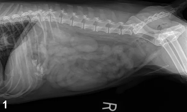
Thoracic/abdominal radiographs: caudal esophageal foreign body (bone opacity); small nonobstructive pieces of bone in stomach, gas-filled stomach, no evidence of aspiration pneumonia or pneumomediastinum, no evidence of gastric or small intestinal obstruction (Figures 1–5).
Figure 1. Right lateral abdominal view
Packed cell volume/total protein: 51%/6.6 g/dL (ranges, 36–60/5.0–7.4)
Figure 2. Ventrodorsal view of abdomen
Blood analysis: Blood urea nitrogen, glucose, electrolytes, and acid–base status within normal limits
Figure 3. Right lateral view of thorax and abdomen
Figure 4. Right lateral thoracic view
Diagnosis
Esophageal foreign body with nonobstructive gastric foreign material
Esophagitis
Figure 5. Ventrodorsal view of thorax
Treatment
PlasmalyteA* with no additives (33 mL/hr IV)
Buprenorphine (0.09 mg IV)
Patient was anesthetized for esophagoscopy with diazepam premedication (in conjunction with previously administered buprenorphine) and intravenous propofol induction. Patient was intubated and maintained on isoflurane and received IV fluids at 10 mL/kg/hr during the procedure. Patient did well under anesthesia and recovered smoothly. He was given famotidine (10 mg IV) and buprenorphine (0.09 mg IV) upon recovery.
Esophagoscopy revealed a bone foreign body 32 cm distal to the canine teeth that was grasped and removed with 3-prong graspers. The bone piece was approximately 2 to 3 cm long and had several sharp points. There was multifocal moderate mucosal damage with 3 small focal areas of white necrotic-appearing tissue. The mucosal damage extended to 37 cm distal to the canine teeth but was not contiguous. The affected area of the esophagus remained mildly dilated after the bone was removed. The stomach contained a moderate amount of fluid. All larger pieces of bone within the stomach were removed. The gastric mucosa was grossly normal.
Postsurgical Treatment
Overnight hospitalization
PlasmalyteA (33 mL/hr IV)
Buprenorphine (0.09 mg q6h IV)
Famotidine (10 mg q24h IV)
Sucralfate (0.5 g q8h PO in liquid slurry)
Ampicillin (150 mg q8h IV)
Maropitant (10 mg q24h IV)
Offered water and canned bland diet
Monitored temperature/pulse/respiration q6h, respiratory rate/effort q2h; performed hourly pain score
The patient began regurgitating during the night and was given 2.5 mg metoclopramide IV and started on 1 mg/kg/day metoclopramide CRI. Famotidine was increased to q12h. The owner opted to continue hospitalization for monitoring during the day and took him home that evening.
Home Therapy
Amoxicillin (120 mg q12h × 10 d PO)
Famotidine (10 mg q12h × 10 d PO)
Sucralfate (500 mg q8h × 10 d PO in liquid slurry)
Metoclopramide (3 mg q8h × 5 d PO)
Canned diet in small amounts frequently for 10 days, gradually shifting to normal feeding schedule/dry kibble diet
Limit activity to avoid induction of regurgitation; however encourage controlled leash walks to stimulate GI motility
Recheck with family veterinarian in 1 to 2 days or sooner if the patient will not eat within 24 hours of returning home, is regurgitating more than 2 to 3 times per day, or exhibits excessive salivation, painful swallowing, lethargy, or increased respiratory effort.
Owner was cautioned about the possibility of development of esophageal stricture 2 to 4 weeks after the procedure, which could cause regurgitation, poor appetite, and discomfort necessitating further care.
Outcome
Patient was reevaluated the day after discharge for regurgitation after receiving his medication that morning. He had been eating and drinking well. His owner admitted that it had been over 12 hours between doses of sucralfate and metoclopramide. Injections of famotidine and metoclopramide were administered. The owner elected to take him back home for continued care. The owner stayed in contact and the patient made a full recovery with resolution of the regurgitation.
*PlasmalyteA is a multiple electrolyte solution
The Specialist’s Opinion
Gretchen Statz, DVM, DACVECC
For the most part, I think this case was handled well. The main issue was the lack of recognition of the esophageal foreign body (FB) on the original radiographs. Luckily this did not adversely affect the outcome and the dog ended up doing very well. Here are a few thoughts on the case.
Esophageal Foreign Bodies
Esophageal FB should be included on the differential list for any animal presented to the emergency room after known FB ingestion, particularly after bone or rawhide ingestion. Several retrospective studies in the past several years have examined esophageal FBs, complications, and outcomes: Bone was found to be common (29.7% of cases in one study1 and 88.6% in another2). Rawhide was another common offender (29.7% in one study1). Esophageal FBs carry a high complication rate, especially if not removed in a timely manner, and should be treated as an emergency. In one study, 82.5% of dogs developed complications, specifically esophagitis.1 In this study, the dogs with extended anesthetic times or longer duration of clinical signs were more likely to have moderate or severe esophagitis, resulting in longer hospital stays.1 In another study, duration of clinical signs adversely affected the ability to remove the FBs using endoscopy, resulting in surgery.2 Esophageal stricture and perforation are more serious complications. One study showed an esophageal perforation rate of 8% and an esophageal stricture rate of 1%3 while other studies show esophageal perforation rates as high as 17.4%4 and 24.1%.2 Mortality rates were generally low but ranged from 01 to 11.1%.2
History Taking
The history in this case was important but complicated by the fact that the initial vomiting/regurgitation episodes were not witnessed by the owner. Even in cases where the episodes are witnessed, owners are not always aware of the difference. It is important to question the owner thoroughly to try to differentiate vomiting from regurgitation. An accurate history can help to guide the diagnostic plan (ie, chest radiographs instead of or in addition to abdominal radiographs in the case of regurgitation). The history of bone ingestion in this case also turned out to be important. As discussed previously, bone is a common esophageal FB and bone ingestion should prompt further investigation when clinical signs are present.
Radiographic Interpretation
This case illustrates the importance of examining the entire radiograph. It is easy to focus attention on the part of the image that we believe to be important especially in an emergency setting with the frequent rush to get to the next case. Every radiograph should be viewed in a systematic manner to be sure all organ systems are evaluated. A written evaluation including normal body systems might be helpful to avoid overlooking a portion of the film. Having the radiographs read by a radiologist made a big difference in this case, exemplifying how this service can be useful in detecting radiographic changes that might otherwise be overlooked.
Antiemetics & Foreign Bodies
This dog was treated with one dose of maropitant prior to discharge. Although maropitant does not have prokinetic effects, it is a potent antiemetic and can mask clinical signs in animals with gastrointestinal FBs, leading to a false sense of security and delaying treatment that can prevent more serious complications (ie, gastrointestinal perforation). In this case foreign material remained in the stomach after numerous vomiting episodes that should have emptied it, so there was risk for the foreign material in the stomach to move into the small intestine and create an obstruction. Despite the potential risk, I think one dose of maropitant was reasonable to keep the dog comfortable overnight as long as follow-up radiographs were part of the plan. The owners were instructed to return to their primary care veterinarian in the morning for repeat films to assess the position of the gastric foreign material. Unfortunately, follow-up radiographs were not performed.
THE GENERALIST’S OPINION
Barak Benaryeh, DVM, DABVP
The successful outcome in this case was due to good care and diligence. The error made in the initial readout of the radiographs was an oversight that can happen to anyone. Two factors helped save this dog: First, the owner was instructed to follow up in the morning and ask for a second set of films to be taken. Second, the radiographs were reviewed by a radiologist.
Reading Radiographs
Although we all learned to “read the entire film,” it is easy to concentrate on one area of the image. This case is a good reminder to look at all areas of a radiograph, even those that are not part of the problem at hand. If you have not already, develop a systematic approach to reading every image. One common system is to work from the periphery of the film to the center. Establish checklists for each body system and tick them off mentally so that every image has been subjected to the same careful reading. Having a radiologist review radiographs can be of great benefit and can significantly reduce errors, as demonstrated in this case.
Follow-up
The instruction given the owners to follow up the next day is an ideal safeguard for patients. No test is perfect and no clinician is above missing an element of a case. Good instructions to your clients about what to look for and when to follow up are critical to continued success as a practitioner. Be sure to have systems in place for telephone follow-up by you or your staff. Explaining to your clients that all tests, especially radiographs, have limitations will help them understand that they need to follow up as well.
Additional Testing
The owners in this case were offered a pancreatic lipase immunoreactivity (PLI) assay as well as a gastrointestinal polymerase chain reaction (PCR) panel, both of which were declined. The dog had a history of eating inappropriate items such as organ meats and bones. The trouble with a PLI in cases such as this is that an elevated level can be misleading: it does not rule out a foreign body and may lead to erroneous treatments for pancreatitis when that is not the primary problem. A gastrointestinal PCR panel is more appropriate in cases of chronic diarrhea and would not have lent much information to this particular case. If recommending tests, it is best to choose ones that will alter your course of action in a meaningful way.
Vomiting vs Regurgitation
When this dog was initially presented, the vomiting was unwitnessed. It was only at the second presentation that the owners had seen the dog retching. To the vast majority of owners, there is no difference between vomiting and regurgitation. They will nearly always call what they see vomiting. History and physical examination can help distinguish between the two. If you have any concern that your patient is regurgitating rather than vomiting, be sure to take thoracic radiographs.
It is fortunate that this dog did well. Esophageal foreign bodies can be difficult, leading to significant complications. This case serves as a good reminder to methodically read a film and protect your patients and yourself by having backup systems in place for follow-up and radiograph review if possible.